Eli G. Cytrynbaum, Clayton M. Small, and Charles B. Kimmel, authors of a new study in Evolution Letters, explain how their discovery of the “extended osteoid” – a nonmineralised bone matrix – influences our understanding of morphological evolution in bony fish.
Sculpins are benthic fish in the superfamily Cottoidea famed for their boniness. Often a bane of sport fishers, sculpins sit on the bottoms of streams, lakes, tidepools and even the open ocean and steal bait – only to be too much of a bottom dweller to offer a good fight and mostly too spiny to make a good meal. With great camouflage and large spines, they deter many a prospective predator and have, within a relatively recent evolutionary time frame, undergone a massive radiation as they have adapted to the range of habitats they now occupy. This relatively rapid diversification in both morphology and life history makes sculpin a great model in which to explore the dynamics of evolution, similar to the famed cichlids of eastern Africa. We have discovered that one mechanism by which these fishes full of prickles and hard bits have been able to achieve such rapid shifts in morphology may be related to a distinctly soft modification: sculpins and a few of their relatives exhibit targeted lack of mineralization in certain bone tissues, allowing for fine tuning of bone morphology without changing the recruitment or placement of bone cells during development.
Bones serve as the scaffolding upon which the rest of the body takes form. As a result, bone shape and size constitute fundamental components of overall morphological diversity in these organisms. Many of the mechanisms which produce variation in bone shape on an evolutionary scale are based on differences in the placement of osteoblasts, bone progenitor cells responsible for the formation and mineralization of bone matrix to produce rigid bone. Changes in the spatiotemporal placement of osteoblasts can be accomplished via controlling the proliferation of osteoblasts, their migration from place to place, or even by converting other cell types to produce bone matrix and has been observed in models ranging from zebrafish to mice and beyond.

Along with altering preliminary bone matrix morphology, we found that sculpins and some related fish employ a completely different strategy to modify bone shape. They produce significant morphological variation by leaving specific swaths of bone matrix nonmineralized, to turn what would have been rigid bone into a floppy membrane.
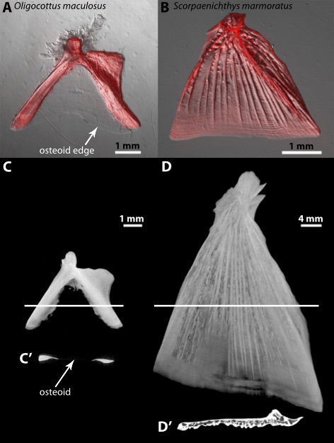
Bone matrix first develops as a flexible, nonmineralized membrane – referred to as osteoid. The membrane – rarely more than a few cells thick – is present on the growing surface of a bone and produced by osteoblasts that are recruited or proliferating. In most species, and in most sculpin bones, osteoid lasts for a vanishingly brief period of time before mineralizing and thus becoming the rigid material we associate with bone tissue. Certain disorders reduce the extent to which bones mineralize, and some mutations or the lack of key nutrients have even been found to stop mineralization throughout the body. However, in many species of sculpins, a region on the bones of their gill cover, in particular a bone known as the opercle, the osteoid remains nonmineralized as it grows. By fine tuning mineralization and bone development carefully enough to leave a variable patch of bone matrix nonmineralized within a larger bone where the rest of the tissue is normally mineralized, sculpins have been able to achieve drastically varied bone morphology, seemingly without altering the spatiotemporal distribution of osteoblasts.

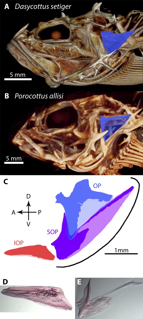
This nonmineralized bone matrix – which we dubbed “extended osteoid” for the manner in which it extends the bone developmental stage of osteoid both spatially and temporally – appears to different extents in various sculpin lineages. In certain species the entire main opercular bone is mineralized, giving it a fan shape, while in other species extended osteoid composes the entire middle portion, making the bone a rigid fork with a flexible sail in between. We observed the fork phenotype in all surveyed freshwater and intertidal sculpins along with some subtidal marine species, and the extended osteoid tissue responsible for this “forkiness” is a major contributor to the morphological variation of the bone.
Opercular extended osteoid gives us a wonderful model for studying evolutionary dynamics, especially as it appears to have arisen several times independently across the phylogeny. Moreover, it provides an opportunity to explore the mechanisms underlying bone development and mineralization as typical osteoid is so small and transient.
Many questions still remain, such as what selective agents may have led to the repeated evolution of this phenotype and what pathways are involved in the maintenance of extended osteoid during development. This novel material holds great potential for further research, along with helping to explain the incredible morphological diversity we observe in sculpin craniofacial bones.
Eli G. Cytrynbaum, Clayton M. Small, and Charles B. Kimmel are all researchers at the University of Oregon. The original paper is freely available to read and download from Evolution Letters.
